Eurostars · Biotech
NanoLoom: AI-Powered Single-Molecule Analysis of DNA–Protein Interactions
In the Eureka Eurostars-3 project NanoLoom, Emoda leads the development of the A…
View
Building healthcare technology since 2018 Schedule a demo with us
Last updated: · By Emoda Software
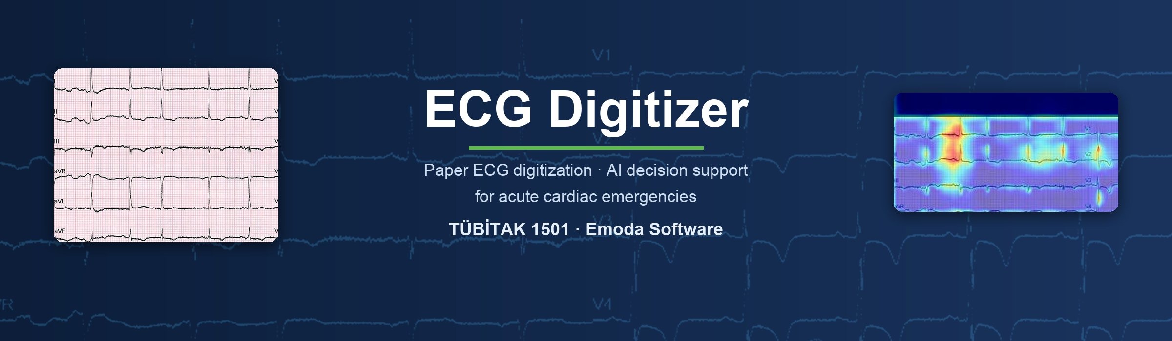
ECG Digitizer (A Technical Solution to Digitize and Analyse Patient ECGs and Accelerate Physician Communication for Faster Diagnosis and Treatment of Acute Cardiac Diseases) was an R&D project carried out by Emoda Software from 1 July 2023 to 31 March 2025 under the TÜBİTAK 1501 Industrial R&D Projects Support Programme, a national funding programme of TÜBİTAK, the Scientific and Technological Research Council of Türkiye. The project delivered an ECG digitization tool that converts printed ECGs into digital signals, a deep learning model that classifies ECG images and a case-based decision support system that compares a new patient with similar past cases. The aim is to shorten the physician's decision time in acute cardiac situations such as chest pain and to digitalise consultation between physicians.
| Project name | A Technical Solution to Digitize and Analyse Patient ECGs and Accelerate Physician Communication for Faster Diagnosis and Treatment of Acute Cardiac Diseases |
|---|---|
| Funding programme | TÜBİTAK 1501 Industrial R&D Projects Support Programme – TÜBİTAK is the Scientific and Technological Research Council of Türkiye, the national public agency funding research and innovation |
| Project no | 3230187 |
| Duration | 1 July 2023 – 31 March 2025 (21 months) |
| Organisation | Emoda Software |
| Clinical data | Anonymised ECG and case data collected, with ethics committee approval, in the emergency department of Çiğli Training and Research Hospital (İzmir, Türkiye) |
| Outputs | ECG digitization tool, ECG labelling software, labelled ECG database, deep learning classification model, case-based decision support system, physician interfaces |
Chest pain is one of the most common reasons for emergency department visits, and in acute coronary syndrome every minute counts. Guidelines recommend that the ECG of a patient with chest pain is recorded and assessed within 10 minutes, and that patients with ST-elevation myocardial infarction (STEMI) reach coronary angiography within 90 minutes. Early diagnosis reduces irreversible loss of heart muscle, so ACS findings such as ST elevation must be recognised fast.
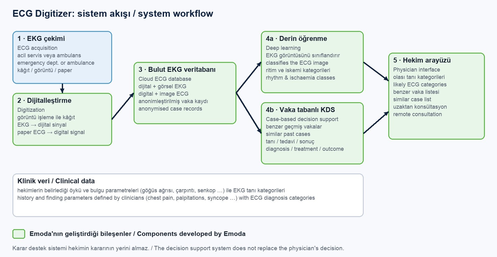
To convert an ECG image to a signal, the scanned or photographed paper ECG is first pre-processed: lighting and contrast differences are corrected, the image is aligned and the relevant regions are separated. The ECG trace is then extracted from the paper grid and converted into a digital signal. The digital ECG is stored both as an image and as a signal, so a patient's previous ECGs are not lost and can be compared and shared between physicians.
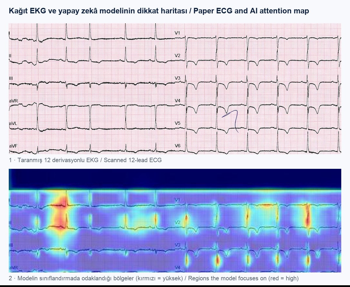
Clinicians on the project labelled the data by assessing the P, Q, R, S and T waves and defined five main ECG classes: tachycardia, bradycardia, rhythms with normal heart rate, other arrhythmias and ECG with signs of previous ischaemia (ST elevation, ST depression, T-wave inversion). A deep learning model was trained on these labelled ECG images. For a new ECG the model gives the probability of each class (e.g. 70% tachycardia, 25% normal heart rate) and shows the regions it focuses on with a heat map. AI-based ECG interpretation is designed as a second pair of eyes that speeds up the physician's assessment.
The case-based decision support system compares a new patient with earlier cases whose diagnosis and treatment are known. The comparison uses history and finding parameters defined by the project's physicians, such as age, sex, chest pain, palpitations and syncope. A machine learning model trained on these parameters predicts the patient's ECG diagnosis category with probabilities and shows the physician the diagnosis, treatment and outcome of similar past cases. According to the report for the second half of 2024, parameters and diagnoses of 1,418 patients had been entered into the system.
In the web interface the physician enters patient information, the ECG file, history and findings and the diagnosis step by step. As soon as the ECG is stored in the cloud it can be sent to other physicians for consultation; the consultation result, diagnosis and treatment are added to the case record, so every new case enriches the decision support database. The goal includes consultation via a mobile app so that ambulance teams can decide quickly even when no cardiologist is available.
After ethics committee approval, ECG data were collected with a scanner provided by the project from patients presenting to the emergency department of Çiğli Training and Research Hospital (İzmir, Türkiye). Personal data were anonymised, and age, sex, symptoms and history were entered through a web interface built for the project. The parameters were defined by the academic physicians on the project.
ECG Digitizer was developed for emergency physicians, general practitioners working in emergency departments, ambulance (112) teams, hospitals that need fast access to cardiology consultation (especially in rural areas) and researchers who want to turn paper ECG archives into digital data for AI studies.
Funding: This project was supported by the TÜBİTAK 1501 Industrial R&D Projects Support Programme under project number 3230187. TÜBİTAK (the Scientific and Technological Research Council of Türkiye) is Türkiye's national agency for funding research and innovation.
ECG Digitizer is a case-based decision support system developed by Emoda with TÜBİTAK 1501 support. It converts paper ECGs into digital signals, classifies them with deep learning and compares a new patient with similar past cases, to speed up diagnosis and consultation in acute cardiac emergencies.
The project was supported by the TÜBİTAK 1501 Industrial R&D Projects Support Programme under project number 3230187 and ran from 1 July 2023 to 31 March 2025. TÜBİTAK, the Scientific and Technological Research Council of Türkiye, is the country's national research and innovation funding agency.
The scanned or photographed ECG image is pre-processed (lighting and contrast correction, alignment, region separation), then the ECG trace is extracted from the paper grid and converted into a digital signal. The digital ECG is stored in the cloud as an image and as a signal.
The model recognises five main classes defined by clinicians: tachycardia, bradycardia, rhythms with normal heart rate, other arrhythmias and ECG with signs of previous ischaemia (ST elevation, ST depression, T-wave inversion). It gives a probability for each class.
It compares the new patient's history and finding parameters, such as age, sex, chest pain, palpitations and syncope, with earlier cases whose diagnosis and treatment are known, and shows the physician the likely ECG category and the diagnosis, treatment and outcome of similar cases.
AI can classify an ECG image into the categories it has learned and show the physician likely rhythm and ischaemia findings with probabilities. In ECG Digitizer this interpretation is used as decision support, while the diagnosis and treatment decision stays with the physician.
The system was designed so that an ECG recorded in the emergency department or an ambulance is stored in the cloud and sent to the physician and to other physicians for consultation, enabling a fast assessment even when no cardiologist is present.
No. ECG Digitizer gives the physician decision support and fast consultation; the diagnosis and treatment decision remains with the physician.
ECG data were collected with ethics committee approval in the emergency department of Çiğli Training and Research Hospital (İzmir, Türkiye) and anonymised. According to the report for the second half of 2024, parameters and diagnoses of 1,418 patients had been entered into the system.
In the Eureka Eurostars-3 project NanoLoom, Emoda leads the development of the A…
View
Emoda is the only participant from Türkiye in the Horizon Europe LUCIA project,…
View
In VILLAGE, funded by the Horizon Europe MSCA Staff Exchanges programme, Emoda d…
View